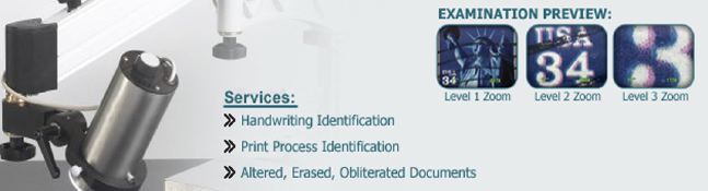
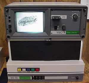
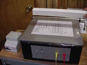
A special instrument for document examination is a UV/IR spectral comparator which makes use of electromagnetic wavelenths and various filters to differentiate inks and reveal hidden material on documents. The Foster & Freeman VSC-4c has a full compliment of visible, UV and IR sources, source and camera filters, coaxial light, transmitted light, oblique light and other features that allow the document examiner to see beyond what the human eye can see. For example, was an addition made to a medical record? Similar spectral comparators are made by Projectina and ACO Electronics (sold here by QDEWill)
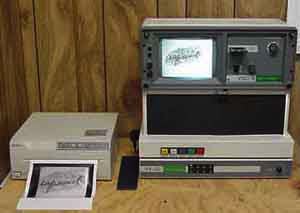
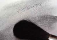
In the demonstration below, a word written with one black pen has been obliterated by scribbling with another black pen. Although the ink all looks the same to the human eye, exciting the document with wavelengths from above and/or below the visible range (IR and UV) and filtering the wavelengths that are reflected to the camera enable the document examiner to see what was previously invisible. The monitor shows that the word "infrared" is beneath the scribble. An image of the monitor's screen can be captured with a digita camera. This is great for inserting images into reports and making court exhibits.
For another example of work with the VSC, visit "Document Examination Fun at Tax Time" - Restoring faced writing on cash receipts.


Opinions reached in the forensic examination of documents and handwriting often hinge on characteristics that are small in size, but large in significance. The examiner must be able to see,
record and evaluate the evidence. Evaluation of evidence calls upon the knowledge, experience, and judgment of the examiner, but first, to see the evidence and record it the examiner needs tools. The
most important tool a document examiner has, aside from his or her own vision and thought process, is the microscope, which helps the examiner to see and record more than is apparent to the unaided eye. Technology brings document examiners new devices that supplement or work with the microscope to accomplish these tasks. But the microscope itself is still essential in document examination.
A microscope that captures two distinct images (one viewed by each eye) which the human brain
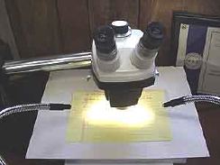
integrates into a three dimensional view of the subject, is a stereomicroscope. A great microscope for document examination is a relatively low power microscope that is both reflective and stereoscopic. Another plus of the stereomicroscope is the natural, comfortable feel of the instrument that results from being able to use both eyes for viewing. The document examiner is often looking through the scope for long periods of time while considering the various details revealed. Comfort becomes important.
In the QDEWill laboratory, handwriting is examined with a low power (7-25x) American Optical
Comparison stereomicroscope. You can visit the microscope page to learn more about this type of
scope and try your skill in identifying microscopic images.
In other situations, such as a case in which the examiner wants to identify the process that produced a document, or to look at a "line sequence" problem (which line was written or printed first), a higher magnification allows a closer look at the "dots" of ink or toner that are on the document in order to make these determinations. For this type of work, I use this Bausch and Lomb stereozoom microscope with lenses and eyepieces providing a range of magnification from 8-160x.&
Sometimes special situations call for special instruments, such as an electrostatic detection device which is used to detect indented writing. I use a VacuBox which consists of a metal box with a porous metal plate at the top. The document is positioned on the plate and covered with a clear protective sheet. A vaccuum is drawn, holding the document tightly to the metal plate, and an electrostatic charge is transferred to the document.
Next, a special toner powder is sprinkled over the protected document and the toner is drawn into the indentations on the document, making them visible. If indentations are revealed the results can be photographed, and the document is usually not harmed.
Perhaps the reader wonders about the source of some pictures presented on this website. The
most useful of all stereomicroscopes has a third "tube" to allow for the attachment of a camera. Typically, a shunt transfers the image from one of the two ocular tubes to the third, or photo, tube. That image would not be a stereo image. In the past, the camera would probably have been a 35mm still camera or a video tape camera. Today, it could easily be a digital camera. Both analog and digital cameras have been the source of images on this website.
Two typical setups are shown below. On the left is a 35mm camera coupled to the microscope. On the right is a Ken-A-Vision 7600 Flex Camera which captures a digital video image through a microscope eyepiece and connects to computer where the image is displayed, captured as a still image, examined, stored, and used in illustrated reports and demonstrative exhibits.
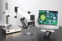
The Viewpoint digital microscope combines optics for magnification with digital technology for image display and capture. Now the examiner can see the big picture on the monitor in addition to, or instead of, seeing it through the eyepiece. These devices supplement, but do not replace, conventional optical microscopes.
Thanks for touring my lab. If you need a document examiner, don't forget the QDE hotline at
1-877-699-7414 or email to ewill@qdewill.com
Copyright © 2001-2008 Emily J. Will All Rights Reserved.